This blog explains what Polysine adhesive microscope slides are and how they improve sample handling in labs.
Standard glass slides work for many lab tasks, but they often fail when tissue sections do not stay attached during staining or washing. This is a common issue in histology and diagnostic workflows, where sample loss can affect slide quality and delay analysis.
To overcome this, Polysine adhesive microscope slides are used to improve tissue grip during processing. These Polysine microscope slides are designed to hold samples firmly, even through multiple steps and heat exposure. With growing demand for reliable slide performance, Blue Star Slides’ products help labs handle advanced applications with greater consistency.
What Are Polysine Microscope Slides?
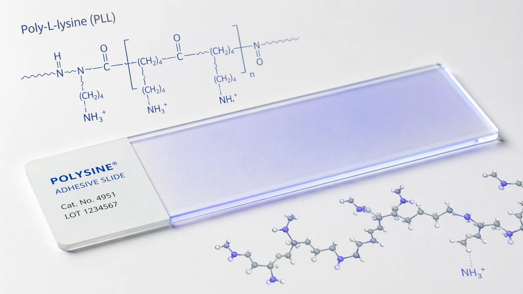
Polysine microscope slides are glass slides coated with a thin, positively charged layer that helps tissue sections stick firmly to the surface. This coating is typically applied to Poly-L-Lysine-coated slides, in which a synthetic amino acid forms a uniform layer across the glass.
The purpose of this coating is simple: to improve adhesion. Unlike plain glass slides, which rely only on surface contact, Poly-L-Lysine-coated slides form a stronger bond with biological samples. This reduces the chances of tissue sections lifting or washing away during processing.
Most Polysine microscope slides also come with a white frosted end, making it easier to label and track samples in busy lab environments. This combination of adhesion and usability makes them a preferred choice in histology and diagnostic work.
How Does the Poly-L-Lysine Coating Work?
The effectiveness of Poly-L-Lysine-coated slides comes from a simple interaction between the slide surface and the biological sample.
- Surface Charge Interaction
Most tissue sections and cells carry a slight negative charge on their surface. The coating used in Polysine adhesive microscope slides is positively charged. When the sample is placed on the slide, this difference in charge creates a strong attraction, helping the tissue attach firmly.
- Strong Adhesion During Processing
Once attached, the sample remains stable through different lab steps. Unlike plain slides, Poly-L-Lysine-coated slides do not need additional adhesives or albumin to keep the tissue in place.
- Stability in Advanced Procedures
Slides may undergo several rounds of washing, staining, and heating in some laboratory methods. Polysine adhesive microscope slides have been developed to cope with such circumstances. These slides are appropriate for both fresh tissue specimens and formalin-fixed paraffin-embedded specimens. They will not be damaged even during high-temperature steps such as antigen retrieval.
Such a regulated adherence process helps make the slides useful in sample retention methods.
Where Are Polysine Adhesive Microscope Slides Used?
Polysine adhesive microscope slides are preferred for applications requiring strong tissue or cell adhesion across multiple processing steps.
- Immunohistochemistry (IHC)
This is one of the most common uses. Slides go through repeated staining and heat-based antigen retrieval. Poly-L-Lysine-coated slides help tissues stay intact throughout the process.
- Histopathology
Used for routine tissue sectioning and staining, such as H&E. These slides reduce the chances of thin sections lifting during preparation and analysis.
- Cytology
In cell smears and FNAC samples, proper adhesion is important for even cell distribution. Polysine microscope slides help keep cells fixed during handling and staining.
- Fluorescence and Confocal Microscopy
These techniques involve multiple wash steps. Strong adhesion from Polysine adhesive microscope slides prevents sample loss during repeated processing.
- In Situ Hybridization (ISH / FISH)
Used in molecular diagnostics, where slides undergo complex chemical treatments. Stable attachment is critical for accurate results.
- Research Laboratories
There may be applications, such as in oncology and neurobiology, where precise and consistent outcomes are ensured by firmly holding samples.
In all these cases, Polysine microscope slides perform better than plain glass slides, where standard adhesion is not enough.
Polysine Slides vs. Plain Glass Slides — What’s the Difference?
When choosing between standard and coated slides, the main difference lies in how well the sample remains attached during processing. Here’s a simple comparison to understand where Polysine microscope slides offer an advantage:
| Feature | Plain Glass Slide | Polysine Adhesive Slide |
| Surface Coating | None | Poly-L-Lysine-coated slides |
| Tissue Retention | Moderate | High |
| Suitable for IHC | Limited | Yes |
| Heat / Antigen Retrieval | Risk of sample loss | Stable during processing |
| Overall Use | Basic lab work | Advanced lab procedures |
| Cost | Lower | Slightly higher |
Plain slides are suitable for routine and educational use where processing is minimal. However, in procedures involving multiple steps, staining, or heat, Polysine adhesive microscope slides offer greater reliability by keeping samples firmly attached.
Choosing the Right Polysine Slides and Where to Source Them
Selecting the right Polysine microscope slides is not just about availability, but about consistency and performance in lab conditions.
- Coating Quality
The Poly-L-Lysine layer should be evenly applied. This ensures uniform adhesion across the slide and reduces the risk of tissue lifting.
- Glass Clarity and Finish
A clear, defect-free surface helps maintain proper visibility. Smooth edges and clean finishing also support safe handling.
- Batch Consistency
Slides should perform the same across different lots. This is important for labs that rely on repeatable results.
- Compliance and Standards
For diagnostic use, it is important to verify that slides meet the required quality certifications and standards.
- Reliable Sourcing
Many labs prefer working with established microscope slide manufacturers in India for a steady supply and consistent quality.
Choosing well-made Polysine adhesive microscope slides helps maintain accuracy and reduces errors during routine and advanced laboratory work.
Why Polysine Slides Are Essential for Advanced Lab Work
In procedures where samples are stained, washed, and heated multiple times, standard slides often aren’t adequate. Polysine adhesive microscope slides provide the added support needed to keep tissues and cells firmly attached, reducing the risk of sample loss during critical steps.
From IHC and histopathology to molecular diagnostics, Poly-L-Lysine-coated slides help ensure consistent, reliable results. This makes them an important choice for laboratories that handle advanced and sensitive workflows.
With growing demand for precision in diagnostics and research, solutions from Blue Star Slides support labs with dependable slide quality. Known for being consistent and reliable, the brand helps labs stay accurate in different tests.
Choosing the right Polysine slides leads to better results and easier processing.